Key facts about age-related macular degeneration
Age-related macular degeneration (AMD) is an eye condition that affects one’s central vision. The condition mainly affects the macula—a part of the retina at the back that regulates central vision. The condition is common among people over 50. People with macular degeneration are unable to perceive objects or people right in front of them, however, they don’t experience a complete loss of vision and can have good peripheral vision. Here are key facts about AMD:
Types
Dry (atrophic) : About 90% of macular degeneration patients are affected by the dry (atrophic) form of the condition. Dry AMD develops when drusen, tiny yellow protein deposits, accumulate under the macula. The deposits make the macula thin and dry. Here, one can experience gradual vision loss, however, most people do not lose their core vision completely. In rare cases, the dry type can give way to the wet form of age-related macular degeneration.
Wet (exudative) : The development of abnormal blood vessels under the retina and macula leads to wet AMD. The vessels can leak blood and liquids, which is a condition called choroidal neovascularization. As a result of the buildup, the macula develops a bulge, leading to dark spots in the center of one’s vision. Wet AMD can quickly turn into a complete loss of vision.
Causes
The retina, located at the back of the eye, transforms light and images reaching the eye into nerve signals to be delivered to the brain. A part of the retina called the macula makes the vision sharper and more precise. The macula, a yellow spot in the middle of the retina, contains high amounts of two natural pigments, lutein and zeaxanthin. When the vessels that supply blood to the macula are damaged over time, it causes age-related macular degeneration. This development also harms the macula. The weakening and thinning of blood vessels along with drusen deposits, are signs of dry AMD. Almost all macular degeneration patients first experience the dry form, and the rest 10% are affected by wet AMD. The condition can be brought on by a family history of macular degeneration. Age and unhealthy lifestyle choices are risk factors for AMD.
Symptoms
Initially, people affected by AMD might not experience any symptoms and can begin to notice problems in their central vision as the condition worsens.
Symptoms of dry AMD: Vision blurring is the most typical sign of dry age-related macular degeneration. Colors and objects near the center of the field of vision can become faded and distorted. One might have trouble seeing fine details or reading text but can easily carry out everyday tasks. One may require more light to read as the condition worsens. The center of vision becomes increasingly darker and blurrier, and with advanced forms of the disease, one might not be able to recognize faces until they are incredibly close.
Symptoms of wet AMD: Straight lines seem warped and deformed when affected by the early stages of wet AMD. One’s vision may have a little dark patch in the middle that becomes larger over time.
Central vision loss is a potential complication of both kinds of AMD. So, if one notices this symptom, they should consult an ophthalmologist immediately.
Diagnosis
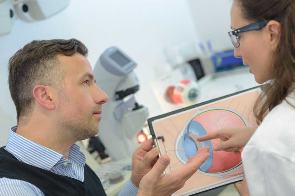
Annual eye exams are crucial in detecting age-related macular degeneration and helping one get timely treatments, as the illness rarely exhibits symptoms in the early stages. The eye doctor examines the retina and macula to look for any changes. One could undergo any of the following tests:
Visual field test : Amsler grids are made of straight lines with a big dot in the middle. The doctor could ask the patient to point out any lines or areas of the grid that are hazy, wavy, or broken. If there is a lot of distortion, one may have AMD, or their vision could be worsening. One can also conduct this visual field test can at home to check their eyesight.
Dilated eye test : Here, eye drops are used to widen the pupils. Then, the healthcare professional uses a special lens to look inside the eyes.
Fluorescein angiography : Here, a yellow dye called fluorescein is injected into a vein of one’s arm. A specialized camera tracks the dye as it passes through the blood vessels in the eye. So, any leakage under the macula can be seen in the photos, letting the doctor know of macular degeneration.
Optical coherence tomography (OCT) : Here, an imaging tool takes precise pictures of the retina and macula placed in the back of the eye. Optical coherence tomography is not an unpleasant or invasive procedure as one can simply look at a lens as the tool takes pictures.
Optical coherence tomography angiography (OCTA) : The diagnostic technique uses laser light reflection instead of fluorescein dye. The OCT then takes 3D images of the blood flow in the eye.
Treatment
Anti-angiogenic treatment options : The doctor injects these prescription options are injected into one’s eyes to prevent the development of new blood vessels and reduce the leakage from abnormal blood vessels that contribute to wet macular degeneration. For some patients, these treatments have been successful in helping them regain vision lost to age-related macular degeneration. On subsequent visits, the treatment will be repeated.
Laser treatment : Eye doctors might advise using high-energy laser therapy to eliminate actively growing abnormal blood vessels.
Photodynamic laser therapy : Here, a light-sensitive treatment option is used as part of a two-step procedure to target abnormal blood vessels. The prescription option is injected into the bloodstream and absorbed by the abnormal vessels. Then, the doctor shines lasers in one’s eye to activate the prescription option and damage the abnormal blood vessels.
Low-vision aids: Devices with unique lenses or electrical systems that can magnify images of adjacent objects are recommended for people with age-related macular degeneration. Examples include reading magnifiers (handheld or electronic), eyeglasses with special lenses, binocular-equipped glasses, electronic glasses, phone applications, and large print items (phones and clocks).
Regular eye check-ups are essential for maintaining one’s vision and eye health and for detecting issues like macular degeneration in the early stages.


